
SERVICES
I trust the innate capability of the body to heal itself. However, we need to provide the body with the kind of food and nutrition that will actually support and encourage the healing capabilities. Imbalances are often caused by certain micro nutrient deficencies. Once the cause is understood, the imbalance can be corrected. In contrast, symptomatic drugs may help alleviate the symptoms but they do not address the cause of the disorder/disease. Unfortunately, this does not make your body any smarter or better!! Yet, symptoms are indicators which tell us in which direction we need to look. Creating an agile and effective immune system is the key to overcome any developing or existing disease condition and experiencing well-being. That's my focus and core competency!
I use the following observations as the basis for the determination of the micro nutritional status of the individual in order to find the right nutritional supplementation. The Blood Electrification and UVBT described further below are additional procedures that train the immune system in the process of self healing. The Magnetic Resonance Pad helps to increase the cell energy levels and supports their detoxification.
Temperature Controlled Blood Microscopy
This is a very unique kind of microscopy which is extremely informative. If you have been through innumerable blood work and every time things show up 'within normal limit' and yet your disease condition continues, maybe what is needed is to be able to observe your blood and see how it is performing (and what is going wrong in your blood as we speak). Conventional blood work may carry a lot of valuable information in many aspects of disease and pathology. However, it is very much possible that there is considerable loss of information in the process of blood stabilization, storage, centrifuging the sample and most of the time testing it several hours later. What I am suggesting here is observing the blood sample within a few minutes under a controlled environment.
In Temperature Controlled Heated Microscopy a drop of blood is taken generally from the tip of the finger. This sample does not comprise only venous blood (like a conventional lab test), but is a combination of venous, arterial and lymphatic fluids. The lymphatic system is the drainage system of the body and hence it transports major impurities in terms of toxins and infections.
The sample is observed using a customized microscope which keeps the blood alive and active. The blood is observed under different magnifications; the primary focus is on the immune cells and its functionality and performance. The sample is also observed for signs of certain nutritional deficiencies, impurities (plagues, debris) and pathogens including, bacteria, funguses, parasites, amoebas and parasitic eggs. Additionally, the presence of certain immune cells like eosinophils and monocytes may suggest parasitic and viral infections, respectively.
This observation is video recorded and can be reviewed at a later consultation or compared with a recording from a different point of time in order to evaluate the changes.
Below are two videos showing
| normal findings | and abnormal findings. |
Functional Leukocyte Observation
In this observation a drop of blood generally from the tip of the finger is taken. Here the circulatory system makes a U-turn and the blood flow is sluggish. As a result impurities tend to settle and may get into the sample. Also, the sample does not comprise only venous blood (like a conventional lab test), but is a combination of venous, arterial and lymphatic fluids. The lymphatic system is the drainage system of the body and hence it transports major impurities in terms of toxins and infections.
The sample is placed on a "heated stage" microscope that actually is set to body temperature which keeps the blood alive and active. Under approximately 10,000 magnification the primary focus is on the immune cells and its functionality/performance. The sample is also observed for signs of certain nutritional deficiencies, impurities and pathogens including plagues, debris, bacteria, parasites, amoebas and parasitic eggs. Additionally, the presence of certain immune cells like eosinophils and monocytes may suggest parasitic and viral infections, respectively.
This observation is video recorded and can be reviewed at a later consultation or compared with a recording from a different point of time in order to evaluate the changes.
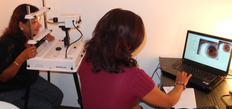
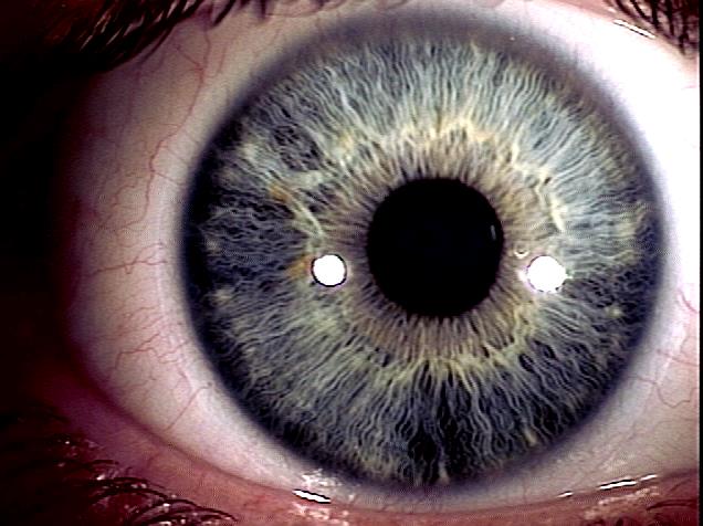
Computer Assisted Iridology
What is Iridology?
Iridology is the science of analyzing the delicate structures of the colored section of the eye, i.e., the iris. The iris is connected to every organ and tissue of the body through the brain and nervous system. The connection is formed by the optic nerve, optic thalami and the spinal cord. From an embryological stand point the iris is formed from the mesoderm and neuroectoderm tissue. It contains sympathetic and parasympathetic flow, very similar to different structures in the body. Thus, the neuro-connections allow the iris to reveal information about inflammation, body constitution, health level and the changes that take place within the body at a cellular level, based on one's life style and life choices.
How is it done?
Computer assisted iridology uses a high definition camera to take pictures of the colored section of the eye. The picture is analyzed by specific software. The software gives a functionality rating for the different organs of the body. Since the interpretation is made by the software, the iridology readings are meaningful and objective, and not just a subjective interpretation of the picture of the iris as in non-computer assisted iridology, which can fall prey to human error.
What is the contribution of Iridology towards assessment of cellular health?
There is an old Chinese proverb which states 'A poor doctor cures; a good doctor prevents'. Iridology is a step in this direction of disease prevention. It is an assessment tool and hence can bring in light the inherent constitutional strength and weaknesses of specific organs. It does not classify body conditions based on disease neither does it diagnose specific diseases. Therefore it cannot be used, for example, to recognize the presence of gallbladder or kidney stones.
Iridology makes it possible to pinpoint inherently weak organs which may or may not be symptomatic or causing any discomfort.
In my practice I am able to break down this test into two levels. One is my interpretation of the iris by analyzing the image of the iris at a macroscopic level. I note the color of the iris; the structure and weave of the fibers (trabeculae). I also look for the presence of defects such as lacunae, lesions, psora, radii solaris etc.
 |
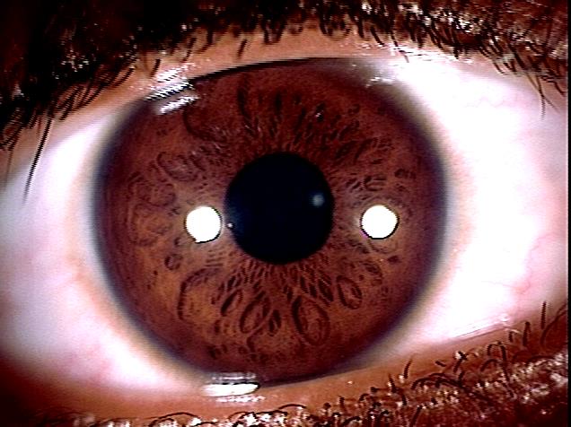 |
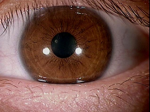
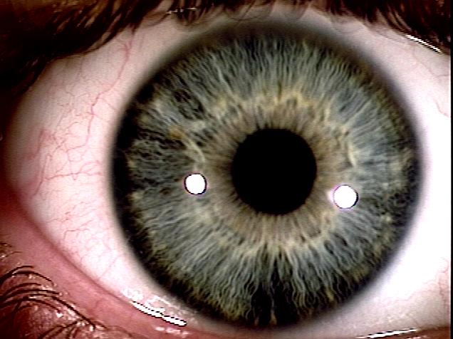
| Eye showing good conditions | Eye showing poor conditions |
There are only two colors of the iris: blue and brown. We also see a combination of true blue and true brown. The other colors that we commonly see are merely deviations from the normal and primarily represent toxin and chemical build up in the body. Below are some images of different kinds of irises and various kinds of anomalies that may be visible even to the naked eye.
I guess after knowing this we may think twice before we admire someone's beautiful sea-green eyes!
 |
 |
| Same eye at first visit (left) and 15 months later (right). The eye has become brighter and more blue. | |
The other part of the test involves understanding and doing a clinical interpretation of the data provided by the software. This is essential for assessing the cellular health of different organs and their functionality
This test is performed during every visit and a track of individual reading is systematically maintained and used as reference to monitor improvement in the patient's health and well-being.
Iridology is the science of analyzing the delicate structures of the colored section of the eye, i.e., the iris. The iris is connected to every organ and tissue of the body through the brain and nervous system. The connection is formed by the optic nerve, optic thalami and the spinal cord. From an embryological stand point the iris is formed from the mesoderm and neuroectoderm tissue and contains sympathetic and parasympathetic flow, very similar to different structures in the body. Thus, the neuro-connections allow the iris to reveal information about inflammation, body constitution, health level and the changes that take place within the body, based on one’s life style and life choices.
For iridology, a high definition picture of the colored section of the eye is taken by a special camera. The picture is digitized and transfered to a computer where a specified software analyzes it. The software gives an effectiveness rating for the different organs of the body. Since the interpretation is made by the software the iridology reading values are meaningful and objective, and not just estimated as in non computer assisted iridology, which can be subject to human error.
Iridology does not classify body conditions by disease, it rather gives important information about tissue health and body constitution. Abnormal readings may suggest a particular organ to be either under-functioning or over-functioning.
- Under-functioning organs may be secondary to under-perfusion due to plaque formation or low nutritional status at a cellular level.
- Over-functioning may be suggestive of infection, trauma and inflammation in that particular organ.
Hair Tissue Mineral Analysis (HTMA)
HTMA is a test performed to measure the mineral content of the hair. The hair sample is prepared in a lab by a series of chemical and high temperature procedures and finally tested for the mineral content using complex equipments. The hair is an ideal tissue for sampling and testing for minerals because:
- As it grows it works like a tape recorder. Important information about cellular metabolic activity gets encrypted in it, thereby providing insight into the body’s biochemical status during the period of hair growth.
- It provides information about the mineral and toxic metal accumulation over a long term and acute exposure.
- It is easy to collect, transport and handle hair sample compared to blood or urine.
- Tests using blood and urine provide information about the mineral level in that moment, almost like a snapshot. Blood levels of a particular mineral may vary based on the diet in that moment. For example eating a banana may temporarily increase the serum potassium level even though the individual may be low in potassium
- Urine levels of minerals actually represent what the body is excreting and may be very different from what is actually retained in the body
- Acute exposure to certain minerals may not show-up in body fluids after a certain time frame. This is because the body tends to shift it into the tissues like the liver, teeth, bones and hair. It is very obvious that out of these different tissues the hair is the easiest to collect and analyze.
- Minerals such as calcium and iron do not show up as low in the serum until they reach critically low levels. HTMA allows us to have a correct estimate of certain minerals in the whole body.
Extensive research is being performed in the field of mineral balance in the body. It is being understood that metabolic dysfunction happen in the body not only because of an excess/ deficiency of certain minerals, but many times it occurs due to an abnormal mineral ratio. An example could be high sodium to potassium ratio which is indicative of early stress, inflammatory reaction or an increased histamine reaction.
Toxic metal exposure is important information as part of the HTMA report. 1980 report of the Environmental Protection Agency stated that human hair can be effectively used as for biological monitoring of key toxic metals. Heavy metals can be inherited by the baby during gestation. HTMA can also detect exposures that occurred years and decades ago. The important heavy metals tested are Uranium, Arsenic, Tin, Mercury, Cadmium, lead and Aluminum.
It is important to understand that unless the exposure to these heavy metals is not on-going or chronic the blood test may not show evidence of elevated level. The reason for this is that as part of the protective action of the body the heavy metal may be moved from the blood into other soft tissue of the body such as the liver, bones, and hair.
Blood Electrification
This is one of the processes we use to stimulate the immune system. A small electrical unit is generally wrapped around the wrist. As the blood flows under the unit any living protein material (e.g. virus, bacteria, parasites etc.) get electrocuted. The dead protein material acts like a powerful immune stimulant.
Further information about Blood Electrificationcan be found here.
Ultraviolet Blood Therapy (UVBT):
The process of Ultraviolet Blood Therapy involves passing blood through a quartz tube surrounded by ultraviolet light and leading it back into the body. This increases the resistance of the patient to a disease. It does this through the following means:
- Ultraviolet light energizes the biochemical and physiological defense system of the body. It does this by partly inducing ozone formation from the oxygen in the blood.
- Ultraviolet light has long been known to inactivate viruses while preserving their ability to be used as antigens in the preparation of vaccines (Levinson, 1945). The proposed theory is that viral genome is more UV-damage sensitive than viral surface antigens. Thus, the virus can be killed by damaging its nucleic acids while, at the same time, leaving antigenic surface components (proteins, glycoproteins, and/or fatty acids) relatively intact. The surface protein acts as an immune-stimulant hence acting as a personalized auto-vaccine.
- UV has been found to be a useful tool in the preventive treatment of platelet-concentrate infusion-induced alloimmunization reactions (Sherman et al. 1991; Pamphilon & Blundell, 1992), and for the prevention of graft-versus-host reactions in transplantation (Leitman, 1989; Kapoor et al. 1992). Here the principal mechanism is thought to be the sensitivity of lymphocytes (that typically contaminate platelet concentrates and carry the HLA antigens responsible for the reactions) to UV inactivation compared to the relative insensitivity of the platelets (which lack nuclear material).
- It has had astonishing results in clearing cyanosis in Hypoxic patients
Further information about UVBT can be found here.
References:
Kapoor, N., Pelligrini, A., Copelan, E., Cunningham, I., Avalos, B., Klien, J., Tutschka, P., (1992). "Psoralen Plus Ultraviolet A (PUVA) in the Treatment of Chronic Graft Versus Host Disease: Preliminary Experience in Standard Treatment Resistant Patients," Seminars in Hematology, Vol. 29, No 2: pp 108-112.
Leitman, S., (1989). "Use of Blood Cell Irradiation in the Prevention of Posttransfusion Graft-vs-Host Disease," Transfus Sci, Vol. 10: pp 219-232.
Levinson, S., Milzer, A., Shaughnessy, H., Neal, J., Oppenheimer, F., (1945). "A New Method for the Production of Potent Inactivated Vaccines with Ultraviolet Irradiation," J. Immunol, Vol. 50: pp 317-329.
Pamphilon, D., Blundell, E., (1992). "Ultraviolet-B Irradiation of Platelet Concentrates: A Strategy to Reduce Transfusion Recipient Allosensitization," Seminars in Hematology, Vol. 29, No 2: pp 113-121.
Sherman, L., Menitove, Kagen, L., Davisson W., Lin, A., Aster, R., Buchholz, D., (1991). "Ultraviolet-B Irradiation of Platelets: A Preliminary Trial of Efficacy," Transfusion, Vol. 32, No 5: pp. 402-407.
Magnetic Resonance Stimulating Pad:
The clearest and most elementary indicator of the 80 trillion cells in our body is their resonation on various frequencies. Each organ resonates in its own frequency. Every molecule has its own quantum signature, a frequency at which it resonates. Organs and tissue create their energy using specific and clearly identifiable resonation. External factors like “electro-smog” and other unhealthy lifestyle habits act as invisible energy robbers. This leads to constant and sometimes permanent stress and a state of diminished energy supply to the cells. “Magnetic noise” is generated mostly from electrical devices such as cell phones, television, radios etc. These are abnormal foreign frequencies which have the potential to interfere with the muted resonance of the cells. This causes reduction in the voltage potential. We may conclude that factors like electro-smog and magnetic noise play a significant role in creating constant strain on an organism leading to a cause of chronic diseases.
In contrast, the Magnetic Resonance Stimulation Pad works by generating a pulsating electromagnetic field similar to the Earth. Unlike magnet therapy where the magnetic field is static, on this mat the magnetic field generated is dynamic (pulsating). This plays a key role in raising cell energy levels and detoxification of the connective tissue.